Diagnostic Imaging
Digital Radiography
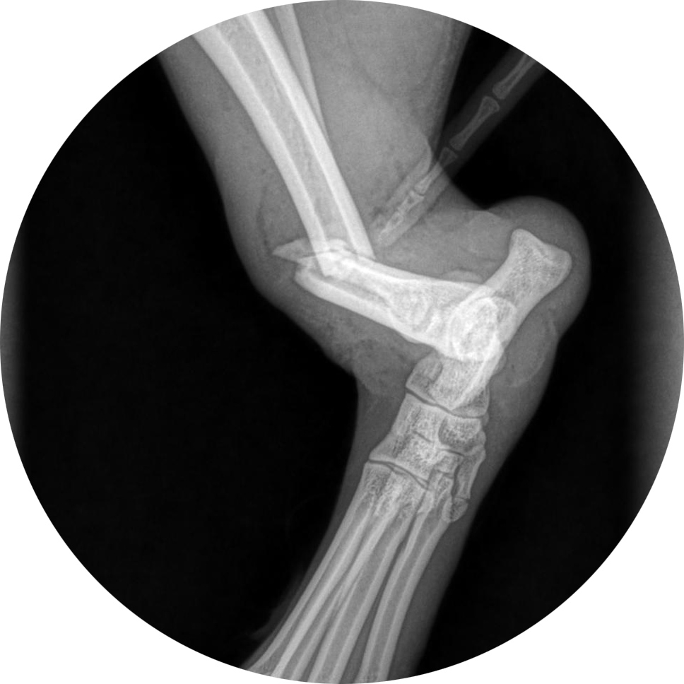
Newburgh Veterinary Hospital has a new, state-of-the-art, digital x-ray machine. Compared to x-rays produced by a traditional machine, the quality of digital radiographs is much better. Because digital radiographs are so much better than traditional x-rays, fewer images are needed in order to make an accurate diagnosis. Digital x-rays are produced quickly and immediately displayed on a computer monitor. They can be manipulated to get a better view of your pet’s bones and internal organs. Our sophisticated digital x-ray equipment produces clear, detailed images that allow our veterinarians to make a more rapid and accurate diagnosis. Radiographs are one of the most important diagnostic tools in veterinary medicine.


Ultrasound
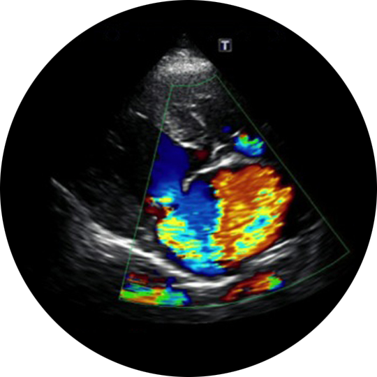
Ultrasound is a pain-free, totally non-invasive technique that uses high-frequency sound waves to produce a real-time moving image of your pet’s internal organs. It allows us a look at your pet’s internal organs, chest and abdomen without surgery or sedation. At Newburgh Veterinary Hospital, we employ ultrasound for a wide-range of diagnostic and medical procedures. It is particularly useful for complete examinations of the abdomen and the heart.
In most cases, an ultrasound procedure is relatively brief. Most importantly, though, an ultrasound, sometimes combined with radiographs, is valuable for making an accurate diagnosis of your pet’s condition and provides effective treatment recommendations.
High Def CT Scanner
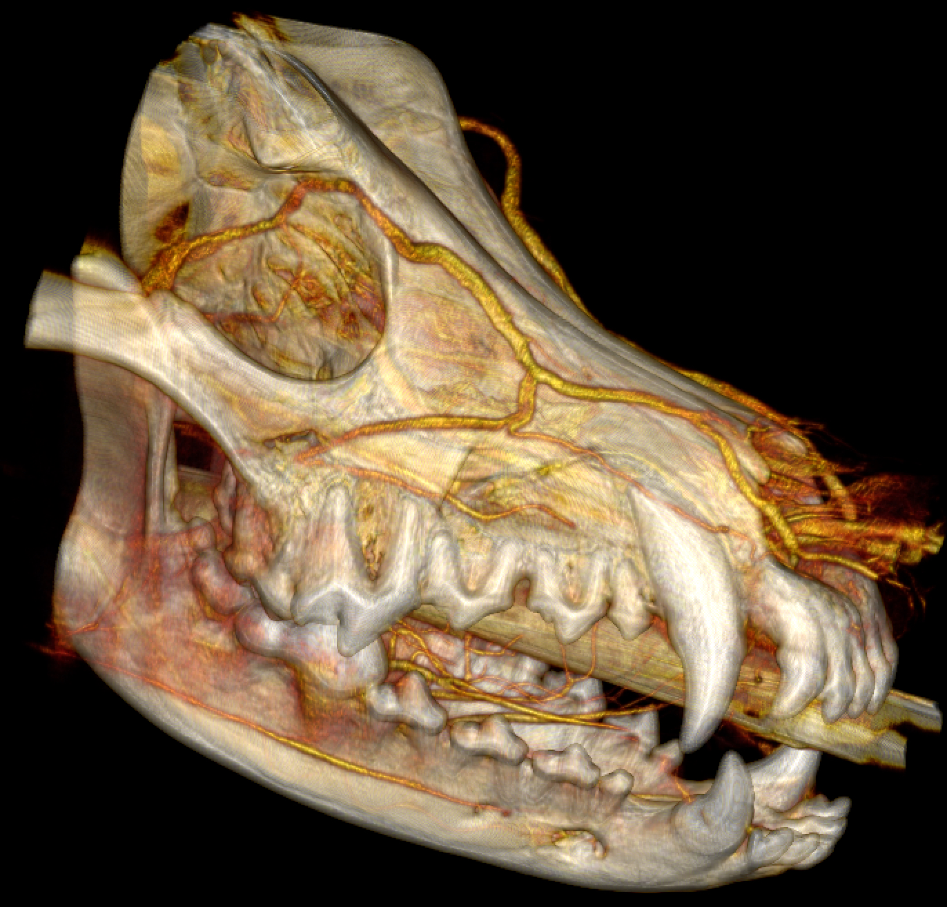
HDVI™ (High-Definition Volumetric Imaging) is a new, proprietary and patented imaging technology, similar to CT (Computed Tomography) that provides unprecedented diagnostic and interventional information for clinicians.
High Def CT Scanning can provide resolution as small as 0.09mm (about the thickness of a human hair). Clinicians can see the data in any angle, thickness or orientation. The results are superior diagnostic confidence – and why HDVI™ is the new standard for primary imaging.
High Def CT Scanning is recommended for:
- Dentistry
- Tumors: identification and assessment
- Trauma
- Foreign bodies
- Discovering abnormalities anywhere in the body
- To assist in surgical planning
Diagnostic Fluoroscopy may be used for: coughing, swallow studies, shunts, collapsing tracheas and more. Interventional Fluoroscopy may be used for: biopsies, endoscopy and more.

